Child Development: From Conception to Birth

A typical pregnancy lasts 270–290 days, or roughly 40 weeks, equivalent to nine months. What transformations unfold within a woman’s body during this time? How does the fetus evolve week by week? What do we understand about the miracle of birth? This article delves into the development of the fetus during the first trimester.
Human embryogenesis is divided into three stages: the initial period (weeks 1–2), the embryonic period (weeks 3–8), and the fetal period, which extends from week 9 until birth. During the initial period, the developing organism is called a conceptus; in the embryonic phase, it is an embryo; and in the fetal stage, it becomes a fetus.
When Does Pregnancy Begin?
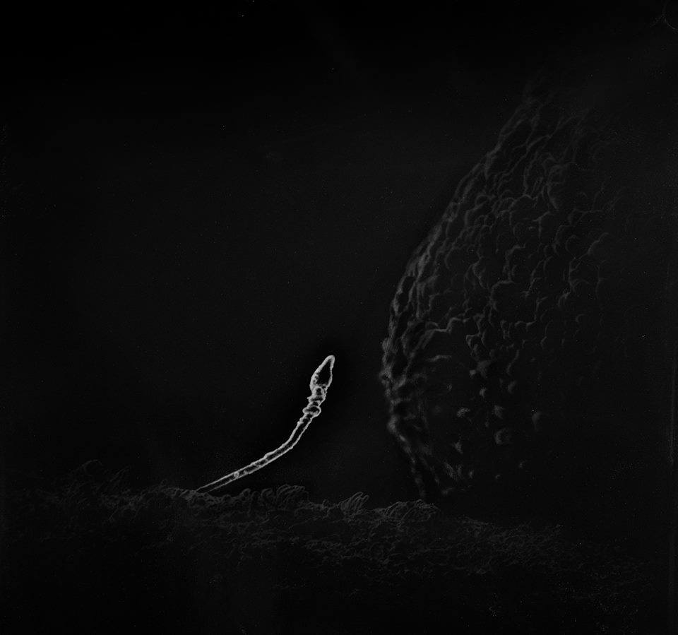
It’s commonly assumed that pregnancy starts the moment fertilization occurs—when a sperm and egg, each carrying a single set of chromosomes, unite to form a zygote, followed by straightforward cell division. However, the process is more intricate. Fertilization takes place in the fallopian tube, after which the zygote travels toward the uterus while simultaneously undergoing cell division. For the first 15 minutes to 32 hours, there may be no changes at all. The zygote divides asynchronously, resulting in an uneven number of cells at times. By days 3–4, it forms a morula—a cluster of 8–12 large, tightly packed cells resembling a mulberry, hence its name.
 Sperm and egg
Under normal conditions, before reaching the uterus, the morula transforms into a blastula, or blastocyst, characterized by an inner cavity and an outer layer of cells. By days 6–7, the blastocyst, now comprising 107 cells, enters the uterus. Having depleted its nutrient reserves and with an increasing demand for energy and oxygen, it attaches to the uterine lining in a process called implantation. At this stage, the conceptus relies entirely on the mother’s body. The walls of the blastocyst burrow into the uterine lining, forming a network of tiny blood vessels that nourish the tiny organism. The embryo itself develops from an inner cell mass, while the outer layer gives rise to the placenta, umbilical cord, and amniotic sac.
Only after the conceptus implants in the uterine wall does pregnancy truly begin.
Week 1
The first week starts with fertilization, progresses through cell division, and culminates in implantation. After insemination and the fusion of sperm and egg, the egg completes its maturation from a secondary oocyte, forming a zygote. This triggers asynchronous, uneven cell division known as cleavage. During cleavage, the number of cells increases, but they do not grow in size; in fact, they lose some mass as energy is consumed. The resulting cells vary in size, with smaller ones forming the outer layer (trophoblast) and larger ones clustering inside (embryoblast). By day 3, the embryo is termed a morula.
On day 4, the outer cells secrete a fluid, creating an inner cavity, and the embryoblast shifts to one pole of the morula, forming a blastocyst. Meanwhile, the uterus prepares to receive the embryo: the uterine lining thickens, releasing nourishing substances like glycoproteins, glycogen, and lipids. The embryo transitions from relying on its internal resources to drawing sustenance from the uterine glands.
Implantation follows, involving adhesion (attaching to the uterine lining) and invasion (penetrating the lining). The embryo releases enzymes that gradually break down the uterine wall, anchoring itself to the epithelium, connective tissue, and eventually blood vessels. Implantation concludes as the uterine lining envelops the embryo, forming a dense network of blood vessels. The embryo now relies on maternal blood for nourishment, marking the shift to hematotrophic nutrition.
Week 2
During the second week, the embryo undergoes primary gastrulation, forming the germ layers that will eventually develop into organs. The embryo becomes a two-layered disc, measuring about 0.25 mm. Extra-embryonic structures—such as the amnion, yolk sac, and chorion—also form to support the embryo’s survival.
Week 3
The third week brings the second phase of gastrulation, resulting in the formation of a third germ layer, the mesoderm. This marks the beginning of germ layer differentiation, laying the foundation for all organs and major functional systems.
The ectoderm gives rise to:
-
Skin
-
Tooth dentin
-
Epithelial linings of the nose, eyes, and ears
-
Nails and hair
-
Nervous system
-
Skull bones
-
Outer eye membranes
Mesoderm differentiation begins around day 20, forming:
-
Notochord (precursor to the spine)
-
Dermis
-
Skeletal, facial, and pharyngeal muscles
-
Bones, cartilage, and blood
-
Lymph, kidneys, blood vessels, heart, and gonads
The endoderm develops into:
-
Lungs
-
Epithelial linings of the stomach, intestines, liver, gallbladder, and pancreas
Starting on day 20, the embryo detaches from extra-embryonic structures, gaining spatial orientation with distinct anterior (future head) and posterior (future pelvis and lower limbs) ends.
Week 4
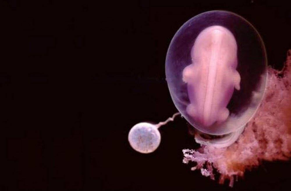
By the fourth week, the embryo floats freely in the amniotic fluid, resembling the embryos of other mammals with a tail and gill-like structures. From day 23, the heart begins to beat, while organ formation continues. The pronephros develops into the primary kidney, brain vesicles form the basis of the brain’s regions, and rudiments of the lungs, duodenum, and liver appear. Small buds on the sides of the body mark the future arms and legs. The mouth, eyes, inner ear, and thymus begin to take shape.
In summary, within one month, the embryo grows to 8 mm, weighs about 3 grams, and develops the rudiments of most systems and organs, with a defined head, limbs, and bilateral symmetry.
 Life in the 4th week
Week 5
The cardiovascular system becomes more complex, developing its conductive pathways. An electrocardiogram at this stage would resemble that of an adult. Major endocrine organs form, and the liver temporarily takes on blood production. The kidneys, lungs, intestines, and nervous system continue to develop. Limb buds evolve into the beginnings of arms and legs.
Week 6
The embryo’s face transforms significantly: the eyes move closer together, the nose develops, and ear structures form. The forebrain and midbrain advance, and limbs begin to show distinct segments (thigh, shin, foot for legs; shoulder, forearm, hand for arms). Red blood cells (erythrocytes) emerge.
Week 7
The seventh week focuses on the skeletal system. Limbs continue to develop, with finger rudiments appearing, though webbing between them persists until week 8. Ossification centers form in the skeleton, a process that continues until around age 25.
Week 8
By the end of the eighth week, lymph nodes form, and gonadal differentiation completes. The embryo now distinctly resembles a human, with the tail disappearing and buttocks forming. Facial features, including lips, become visible, as do pulmonary blood vessels and heart ventricles. The embryo measures 4 cm and weighs 5 grams. The neck develops, and the head transitions from cylindrical to round, revealing the nose and outer ear. Limbs are clearly defined, with visible fingers and segments. Muscle tissue begins to form, and by the week’s end, muscles can contract.
Brain hemispheres continue to develop, with the cortex forming. From day 50, brain activity can be detected as electrical impulses, a key indicator of life.
Transition to the Fetal Period
The embryonic period ends, and the fetal period begins. During the third month, organs continue to form, and some start functioning. The face becomes more defined, and the placenta takes shape.
Week 9
Taste buds appear on the tongue, and the fetus begins to swallow amniotic fluid, suck, and respond to stimuli, reflecting early nervous system development.
Week 10
The fetus’s face is fully formed and mobile. The body becomes sensitive to stimuli like heat, pain, or cold, prompting facial reactions such as grimacing, driven by the maturing nervous system.
Week 11
By week 11, the heart functions normally, beating at 130–150 beats per minute. The intestines are nearly fully formed and begin peristaltic movements.
Week 12
White blood cells (leukocytes) appear in the blood. Hemoglobin is present in small amounts, increasing significantly closer to birth. The skeleton continues to ossify, and fingers and toes become distinct, with the beginnings of fingerprints and nails. External genitalia form but are not yet distinguishable. The vocal apparatus develops, though vocalization is possible only after birth. The fetus weighs up to 25 grams and measures up to 9 cm.
During the third month, the fetus makes its first movements, though these are typically imperceptible to the mother, especially in a first pregnancy.


